
OR
Gastroenterology - Gastrointestinal Infections
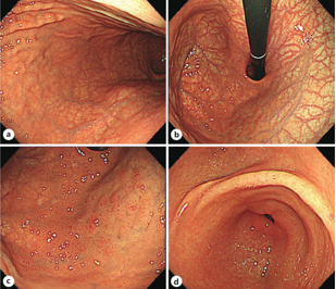
A 40-year-old woman presented with microcytic anemia and underwent esophagogastroduodenoscopy (EGD). EGD revealed extensive mucosal atrophy with prominent submucosal vascularity throughout the gastric corpus, with relative sparing of the fornix. The fornix showed residual oxyntic mucosa with diffuse erythema and pseudopolypoid lesions (Figure 1). Based on these findings, autoimmune gastritis (AIG) with concomitant active Helicobacter pylori infection was suspected. The diagnosis was further supported by serum anti-H. pylori antibody, H. pylori stool antigen, and anti-parietal cell antibody (PCA) testing. The patient received oral iron supplementation and H. pylori eradication therapy. Follow-up EGD and serological evaluation demonstrated regression of gastric mucosal atrophy and partial restoration of the fundic glands following H. pylori eradication, indicating a favorable response to treatment.
© 2026 Imedsource, All Rights Reserved
For unrestricted access to content and a seamless experience,
Complete your profile to help us deliver a more personalized experience.